Cranial Vault Reshaping for the Treatment of Craniosynostosis
A Guide for Parents
Craniosynostosis is a term that most parents are not familiar with—until a doctor mentions it in relation to their own child. The moment you hear the diagnosis, it may feel as if your world has been turned upside down. A rush of emotions floods in—confusion, fear, worry. What does this mean for your baby? Will it affect their brain development? Will they need surgery? How serious is it?
As you start researching, you quickly realize that craniosynostosis isn’t just about an unusual head shape; it’s a condition that, if left untreated, can have serious consequences on brain growth and skull development. One thing becomes clear: The most common and effective treatment is cranial vault reshaping surgery. And suddenly, you’re faced with an even bigger challenge—the thought of your child undergoing major surgery.
This page was written for you—the parent who wants to understand everything about cranial vault reshaping so you can make the best decisions for your child. If your doctor has recommended this procedure, you likely have a million questions swirling in your mind. What does the surgery involve? Is it safe? Will your child be in pain? How long does recovery take? What happens if you don’t do the surgery? What are the long-term effects?
These are all valid concerns, and the goal of this article is to provide clear, detailed, and reassuring information to guide you through the entire process. It is completely natural to feel overwhelmed, but knowledge is a powerful tool. Understanding what to expect can replace fear with confidence and uncertainty with clarity.
Cranial Vault Reshaping for the Treatment of Craniosynostosis
A Guide for Parents

Table Of Contents
What is Cranial Vault Reshaping?
There are many resources available on craniosynostosis, but most are written from a purely medical perspective. While clinical studies and journal articles provide important insights, they can often feel cold and technical, making them difficult for nonmedical readers to digest. This page takes a different approach—it is written specifically for parents.
In this guide for you, the parent:
- We will break down medical terms in an easy-to-understand way without oversimplifying the facts.
- We will focus solely on cranial vault reshaping, rather than overwhelming you with information about all possible treatments for craniosynostosis.
- We will walk you through each stage of the process step by step, from the first diagnosis to long-term outcomes, so you always know what to expect.
- We will address the emotional side of this journey, recognizing that craniosynostosis isn’t just a medical condition—it’s something that deeply affects families.
You are not alone in this process. Thousands of families have gone through this journey, and many have shared their experiences so that others, like you, can feel more prepared and supported.
What Is Cranial Vault Reshaping, and Why Is It Needed?
Cranial vault reshaping is a surgical procedure designed to correct the abnormal skull growth caused by craniosynostosis. In simple terms, it involves carefully removing, reshaping, and repositioning parts of the skull to allow for normal brain development and a more typical head shape.
While every parent hopes to avoid surgery for their child, the reality is that leaving craniosynostosis untreated can lead to serious problems, including:
- Increased intracranial pressure: The skull normally expands as the brain grows, but when the sutures fuse too early, the brain may not have enough space to develop properly. This can lead to pressure inside the skull, which may cause headaches, developmental delays, and, in severe cases, vision loss or cognitive issues.
- Abnormal head shape: While some mild cases may not require surgery, significant skull deformities can impact a child’s appearance and, in some cases, even their ability to wear glasses, helmets, or other headgear comfortably as they grow.
- Potential developmental delays: Some children with untreated craniosynostosis experience speech, motor, or cognitive delays, especially in cases where brain growth is restricted.
- Psychosocial concerns: As a child grows older, a noticeable skull deformity may lead to self-esteem issues or teasing from peers. While appearance is not the primary reason for surgery, it is an important consideration for many families.
Cranial vault reshaping is a highly effective procedure that has helped thousands of children live healthy, normal lives. Thanks to advances in surgical techniques, this procedure is now safer and more successful
than ever before.
But knowing that doesn’t necessarily make it easier to process as a parent, which is why having the right information and support is so important.
What You’ll Learn

Understanding what to expect at every stage can make the process feel more manageable.
This page will guide you through the journey of cranial vault reshaping, covering:
- How craniosynostosis is diagnosed: What signs doctors look for, what tests may be needed, and how to confirm the condition
- Who needs surgery and when: How doctors decide if cranial vault reshaping is necessary, the ideal age for surgery, and different types of craniosynostosis that require treatment
- Preparing for surgery: What tests and evaluations will happen before surgery, how to prepare your child physically and emotionally, and what parents should expect on the day of the procedure
- A detailed explanation of the surgery itself: What happens in the operating room, how surgeons reshape the skull, and what safety measures are in place
- The recovery process: What to expect in the hospital, how long swelling and discomfort will last, and how to care for your child at home
- Long-term outcomes: How your child’s skull will develop over time, potential future interventions, and what long-term follow-up care looks like
- Support for parents and families: How to find emotional support, connect with other parents, and advocate for your child’s care
- Future innovations: The latest advancements in cranial vault reshaping, including 3D surgical planning and less invasive techniques
By the end of this page, you will have a deep understanding of cranial vault reshaping, from diagnosis to long-term outcomes. Most importantly, you will feel empowered and informed as you make decisions about your child’s care.
A Final Word Before We Begin
If you are reading this, chances are that you are feeling a mix of emotions—anxiety, hope, uncertainty, and love for your child. That’s completely normal. Every parent who has gone through this journey has felt the same way.
While the idea of surgery may feel overwhelming right now, you will not be facing this alone. Your child’s medical team, support groups, and other families who have walked this path before you will all be sources of guidance and reassurance.
The best thing you can do for your child right now is to learn, ask questions, and be as informed as possible.
Now, let’s start at the very beginning:
How craniosynostosis is diagnosed and what signs doctors look for.


Diagnosis
When parents first notice something unusual about their baby’s head shape, it can be easy to dismiss it as a normal variation. After all, many newborns have slightly misshapen heads due to their position in the womb or the birth process. In some cases, a baby’s skull may appear uneven due to positional molding, where certain areas flatten from prolonged pressure in one position.
However, when the shape seems persistent or unusual, doctors may begin to suspect craniosynostosis—a condition that requires careful evaluation and, in many cases, surgical intervention.
Understanding how craniosynostosis is diagnosed is an important first step in your journey. A correct diagnosis ensures that your child receives the right treatment at the right time.
This section will walk you through the signs that lead to a craniosynostosis evaluation, the medical professionals involved, the diagnostic process, and the tests used to confirm the condition.
Recognizing the Signs: When Should Parents Be Concerned?
In the early weeks of a baby’s life, parents and pediatricians begin to observe the shape of the infant’s head. While some minor irregularities are normal and may resolve on their own, certain signs may indicate a deeper issue.
Parents should be aware of the following signs of craniosynostosis:
- An abnormal head shape that does not improve over time – Many newborns have a slightly uneven head at birth, but if the shape remains unusual or seems to worsen, it may be a sign of premature suture fusion.
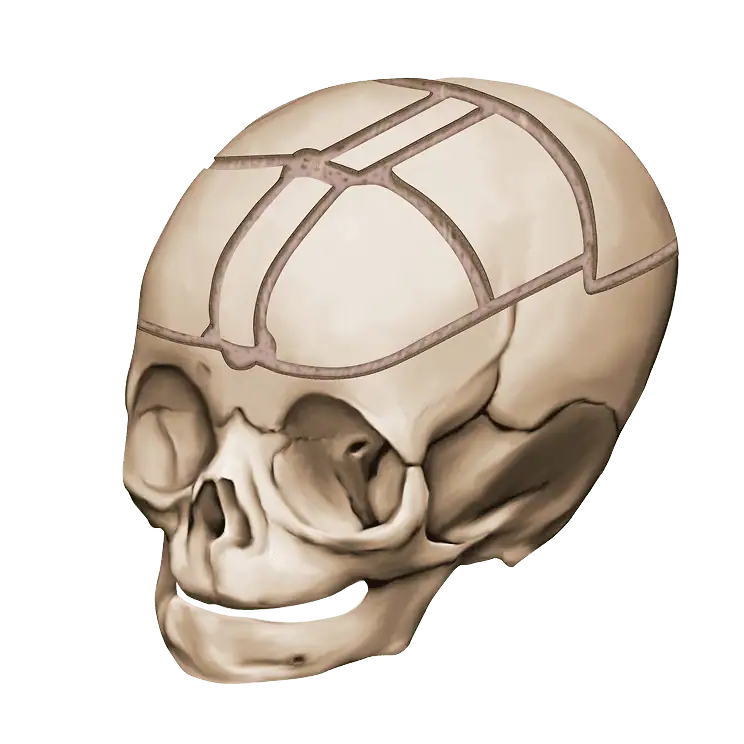
- A ridge or hard line along the skull – In some cases, parents or doctors may be able to feel a firm ridge along the baby’s head where a skull suture has fused too early.
- Asymmetry of the forehead or face – Depending on which suture is affected, the baby’s forehead, eye sockets, or face may appear uneven.
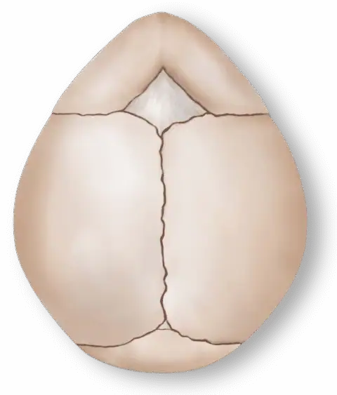
- Lack of the fontanelle (commonly known as the “soft spot”) or early closure – The fontanelle usually remains open for several months to allow skull growth. If it seems unusually small, closes too early, or is completely absent, it could be a sign of craniosynostosis.
- Unusual head dimensions – Some babies with craniosynostosis may have heads that are too long, too wide, or appear triangular due to abnormal skull growth.
- Increased pressure inside the skull (in severe cases) – Although not common in mild cases, untreated craniosynostosis can sometimes lead to symptoms such as irritability, vomiting, developmental delays, or bulging eyes due to rising intracranial pressure.
It’s important to note that not all head shape irregularities mean craniosynostosis. Other conditions, such as positional plagiocephaly, can cause flattening of the skull due to external pressure rather than suture fusion.
Unlike craniosynostosis, positional plagiocephaly can often be corrected with repositioning techniques or helmet therapy.
However, because craniosynostosis requires surgical treatment, early detection is critical. If there are any concerns about your baby’s head shape, a medical evaluation is essential to determine the underlying cause.
Who Diagnoses Craniosynostosis?
When parents suspect something unusual about their child’s skull shape, the first step is usually a visit to the pediatrician. During well-baby visits, pediatricians routinely check head growth and skull shape. If they notice irregularities, they may refer the child to specialists who have more expertise in diagnosing and treating craniosynostosis.
These specialists may include:
- Pediatric neurosurgeons – Experts in brain and skull development who perform surgical treatments for craniosynostosis
- Craniofacial surgeons (plastic surgeons specializing in skull and facial surgery) – Specialists who focus on reshaping the skull and ensuring proper head growth
- Pediatric geneticists (in cases where a doctor suspects the craniosynostosis is part of a genetic syndrome) – Specialists who perform genetic testing
A team-based approach is often used in craniofacial clinics, where multiple specialists collaborate to provide a thorough evaluation and treatment plan.
The Diagnostic Process
Once a doctor suspects craniosynostosis, they will use a combination of physical examination and imaging tests to confirm the diagnosis.
Physical Examination
The first step is a hands-on assessment of the baby’s head shape, skull sutures, and overall development. During this exam, the doctor will:
- Measure head circumference – This enables a comparison with normal growth charts to see if the skull is growing appropriately.
- Feel for suture ridges – A fused suture may form a firm ridge that can be detected by touch.
- Examine the forehead and facial symmetry – Different types of craniosynostosis affect different parts of the skull. The doctor will look for signs of asymmetry, eye socket abnormalities, and other craniofacial irregularities.
- Check for soft spot closure – An unusually small or missing fontanelle may be a red flag for early suture fusion.
In some cases, an experienced craniofacial specialist may be able to strongly suspect craniosynostosis based on the physical exam alone. However, imaging is usually needed for confirmation.
Imaging Tests to Confirm Craniosynostosis
If the doctor suspects craniosynostosis, they will order imaging tests to get a clear picture of the skull. The most frequently used tests include:
- 3D CT scan – This is the gold standard for diagnosing craniosynostosis. A 3D CT scan provides a highly detailed view of the skull, allowing doctors to see which sutures are fused and how the skull shape is affected.
- X-rays (less commonly used today) – In some cases, simple skull X-rays may be taken to check for suture fusion, but they are not as detailed as a CT scan.
- MRI – While not typically used for diagnosing craniosynostosis, an MRI may be recommended if doctors need to evaluate brain structures in addition to the skull.
Because CT scans involve radiation exposure, they are typically used only when necessary.
However, modern technology allows for low-radiation CT scans that provide clear imaging with minimal risk.In most cases, one CT scan is enough to confirm the diagnosis and guide surgical planning.
Finalizing the Diagnosis and Moving Forward
Once the imaging results are reviewed, the medical team will confirm whether craniosynostosis is present and discuss the next steps with the child’s parents.
This conversation will typically cover:
- Which sutures are affected
- How the condition is impacting skull growth
- Whether surgery is necessary and the ideal timing
- What surgical options are available
For many parents, receiving a diagnosis of craniosynostosis can be overwhelming. However, early detection allows for timely intervention, positive surgical outcomes, and low risk of complications. If cranial vault reshaping is recommended, the next step is understanding who qualifies for surgery and when it should be performed, which we will cover in the next section.


Indications and Patient Selection
When a child is diagnosed with craniosynostosis, one of the most pressing questions parents have is whether surgery is truly necessary. Some may wonder if their child’s head shape will improve on its own, while others worry about waiting too long and missing a critical window for intervention. Understanding who needs cranial vault reshaping and when it should be performed is essential for making informed decisions about your child’s care.
This section will explain the factors that determine whether a child is a candidate for cranial vault reshaping, including the age at which surgery is typically performed, the types of craniosynostosis that require surgery, and other considerations that influence treatment decisions.
Why Surgery? When Cranial Vault Reshaping Is Recommended
Cranial vault reshaping is not performed on every child with an unusual skull shape. In some cases, an irregular head shape may be caused by positional plagiocephaly, which can often be managed without surgery through repositioning techniques or helmet therapy. However, when craniosynostosis is confirmed, surgery is usually recommended for the following reasons:
- To allow normal brain growth: The skull expands as the brain grows. If a suture has fused too early, it may restrict skull expansion, leading to increased intracranial pressure in severe cases.
- To prevent complications: In some cases, untreated craniosynostosis can lead to vision problems, developmental delays, or other neurological issues due to restricted brain growth.
- To improve head shape and facial symmetry: While cosmetic concerns are not the primary reason for surgery, significant skull deformities may impact a child’s future self-esteem and ability to wear headgear (such as helmets or glasses).
- To avoid future corrective procedures: Early intervention often leads to better surgical outcomes and may prevent the need for more extensive corrections later in childhood.
The decision to proceed with surgery depends on several key factors, which we will explore in detail.

The Role of Age in Surgical Timing
One of the most important factors in determining when to perform cranial vault reshaping is the child’s age. The timing of surgery is critical because the skull bones are most malleable in infancy, allowing for easier reshaping and faster healing. Age is an important consideration for the following reasons:
Optimal age for surgery:
- The ideal age for cranial vault reshaping is typically between 6 and 12 months of age.
- At this stage, the skull bones are still soft and flexible, making it easier for surgeons to reshape the skull and allowing the child’s natural growth to help maintain the corrected shape.
- Surgery before the child is 6 months old is possible in some cases, especially for endoscopic-assisted procedures, but traditional open cranial vault reshaping is usually performed after 6 months.
- If craniosynostosis is diagnosed late, surgery may still be performed in older infants or toddlers, but the procedure may be more complex due to thicker, less flexible skull bones.
Why early surgery is preferred:
- Better surgical results: Younger skulls are easier to reshape and heal more quickly.
- Lower risk of complications: Early intervention prevents intracranial pressure buildup and allows the brain to grow normally.
- Less noticeable scarring: Scars tend to heal better in infancy and may be less visible as the child grows.
Although surgery is typically recommended before the first birthday, older children can still undergo cranial vault reshaping if necessary. The surgical approach may differ, but successful outcomes can still be achieved.
Types of Craniosynostosis and Their Impact on Surgery
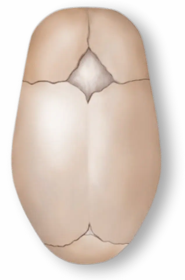
- Sagittal
- Coronal
- Metopic
- Lambdoid
Metopic
- Overview
- This is the most common type of craniosynostosis. It affects the sagittal suture, which runs from the front to the back of the skull.
- It results in a long, narrow head shape (scaphocephaly).
- Surgery is almost always recommended, as this type of fusion can restrict side-to-side skull expansion and lead to intracranial pressure problems if left untreated.
- Cranial vault reshaping is typically performed between 6 and 12 months of age for the best outcome.

The overall incidence of Craniosynostosis is approximately 1 in 2000/3000 births. Metopic synostosis is relatively rare, accounting for between 5% and 15% of all craniosynostosis cases.
Diagnostically, the condition can be identified through a physical examination, where the triangular shape of the forehead and ridging are noticeable. Imaging techniques, such as computed tomography (CT) scans, can confirm the diagnosis and assess the extent of the fusion.
Treatment typically involves surgery to correct the skull shape and allow for normal brain growth. The surgical procedure, usually performed in the first year of life, involves removing and reshaping the fused bones to create a more typical skull contour. Please refer to our Treatment Options page for a more detailed explanation of these surgical procedures.
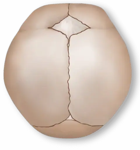
Coronal

- Overview
- This type involves premature fusion of one (unilateral) or both (bilateral) coronal sutures, which run from the ears to the top of the skull.
- It leads to forehead and facial asymmetry if one side is affected and a broad, flat forehead if both sides are involved.
- Cranial vault reshaping is almost always required, as untreated coronal craniosynostosis can affect eye socket development and facial symmetry.
- Surgery is usually performed around 9-12 months of age.
The overall incidence of Craniosynostosis is approximately 1 in 2000/3000 births. Metopic synostosis is relatively rare, accounting for between 5% and 15% of all craniosynostosis cases.
Diagnostically, the condition can be identified through a physical examination, where the triangular shape of the forehead and ridging are noticeable. Imaging techniques, such as computed tomography (CT) scans, can confirm the diagnosis and assess the extent of the fusion.
Treatment typically involves surgery to correct the skull shape and allow for normal brain growth. The surgical procedure, usually performed in the first year of life, involves removing and reshaping the fused bones to create a more typical skull contour. Please refer to our Treatment Options page for a more detailed explanation of these surgical procedures.
Sagittal
- Overview
- This type affects the metopic suture, which runs from the nose to the top of the head.
- It causes a triangular forehead shape (trigonocephaly), with a narrow forehead and widely spaced eyes.
- The need for cranial vault reshaping depends on the severity; mild cases may not require intervention, but moderate and severe cases usually benefit from surgery.
The overall incidence of Craniosynostosis is approximately 1 in 2000/3000 births. Metopic synostosis is relatively rare, accounting for between 5% and 15% of all craniosynostosis cases.
Diagnostically, the condition can be identified through a physical examination, where the triangular shape of the forehead and ridging are noticeable. Imaging techniques, such as computed tomography (CT) scans, can confirm the diagnosis and assess the extent of the fusion.
Treatment typically involves surgery to correct the skull shape and allow for normal brain growth. The surgical procedure, usually performed in the first year of life, involves removing and reshaping the fused bones to create a more typical skull contour. Please refer to our Treatment Options page for a more detailed explanation of these surgical procedures.
Sagittal
- Overview
- This type of craniosynostosis affects the lambdoid suture, located at the back of the skull.
- It causes asymmetry at the back of the head, sometimes mistaken for positional plagiocephaly.
- This is the rarest type of craniosynostosis, but if confirmed, surgery is often recommended.

The overall incidence of Craniosynostosis is approximately 1 in 2000/3000 births. Metopic synostosis is relatively rare, accounting for between 5% and 15% of all craniosynostosis cases.
Diagnostically, the condition can be identified through a physical examination, where the triangular shape of the forehead and ridging are noticeable. Imaging techniques, such as computed tomography (CT) scans, can confirm the diagnosis and assess the extent of the fusion.
Treatment typically involves surgery to correct the skull shape and allow for normal brain growth. The surgical procedure, usually performed in the first year of life, involves removing and reshaping the fused bones to create a more typical skull contour. Please refer to our Treatment Options page for a more detailed explanation of these surgical procedures.
Additional Factors Influencing Patient Selection
Factors in Determining the Need for Surgery
While age and suture involvement are the primary factors in determining the need for surgery, doctors also consider other medical and developmental factors, such as:
- Signs of increased intracranial pressure – If a child has symptoms such as persistent headaches, vomiting, or developmental delays, early surgery may be necessary.
- Family history and genetic syndromes – Some cases of craniosynostosis are linked to genetic conditions such as Crouzon syndrome or Apert syndrome, which may require a different surgical approach.
- Overall health and medical conditions – Children with additional health concerns may need specialized surgical planning and a multidisciplinary team approach.
What if Surgery Is Delayed or Declined?
Some parents may be hesitant about surgery or may need time to process the diagnosis. While cranial vault reshaping is not always an emergency, delaying surgery too long may:
- Make the procedure more complex due to thicker skull bones
- Increase the risk of intracranial pressure problems in some cases
- Lead to less effective reshaping compared with early intervention
If surgery is declined entirely, long-term monitoring is essential to watch for signs of increased intracranial pressure or other complications. However, most cases of significant craniosynostosis require surgical intervention for the best outcomes.


Preoperative Preparation
Once the decision has been made to proceed with cranial vault reshaping, parents often find themselves in a whirlwind of emotions—relief that a treatment plan is in place, anxiety about the upcoming surgery, and a deep desire to ensure they are doing everything possible to prepare their child.
Preparing for cranial vault reshaping involves more than just scheduling a surgery date. It includes medical evaluations, understanding anesthesia, preparing for the hospital stay, and emotional readiness for both your child and your family. Knowing what to expect in the weeks leading up to surgery can ease anxiety and help you feel more in control.
This section will walk you through:
- Medical tests and preoperative evaluations
- Understanding anesthesia and surgical risks
- Practical preparations for surgery day and your hospital stay
- Emotional support for you and your child
Medical Evaluations and Preoperative Testing
Before cranial vault reshaping surgery, your child’s medical team will perform a series of evaluations to ensure your child is healthy enough for the procedure. These assessments help identify any underlying conditions that may need to be managed before surgery and allow the surgical team to plan the safest possible approach.
Preoperative Appointments: Who Will You Meet?
You will likely have multiple appointments leading up to the procedure. These appointments may involve the following medical team members:
- Craniofacial surgeon and/or neurosurgeon – They review the surgical plan, answer your last-minute questions, and ensure that you fully understand the procedure.
- Pediatrician – They assess your child’s overall health and ensure there are no underlying infections, colds, or other illnesses that could delay surgery.
- Anesthesiologist – They discuss the anesthesia process, how your child will be monitored during surgery, and any risks involved.
- Preoperative nurse or hospital coordinator – They provide instructions on fasting before surgery, what to bring to the hospital, and any necessary paperwork.
Common Preoperative Tests
Several tests may be ordered to ensure your child is in good health before undergoing surgery. These typically include:
- Blood tests – These tests are performed to check hemoglobin levels, blood clotting ability, and overall health markers.
- Imaging (CT or MRI, if needed) – While a 3D CT scan is often done during diagnosis, additional imaging may be ordered to assist with surgical planning.
- Cardiac evaluation (if needed) – If your child has a heart condition or any medical concerns, a pediatric cardiologist may evaluate them to ensure their heart can safely handle anesthesia.
If any issues are detected, such as low hemoglobin (which could increase the risk of excessive bleeding),
your child’s surgical team may adjust the timing of the procedure or recommend treatments to correct the issue before surgery.Understanding Anesthesia and Surgical Risks
One of the biggest concerns parents have is anesthesia—the idea of their child being put in an induced sleep for a major surgery can be frightening.
However, modern pediatric anesthesia is extremely safe, and cranial vault reshaping is performed under the care of highly specialized pediatric anesthesiologists.
How Anesthesia Works for Cranial Vault Reshaping
To ensure your child’s safety and comfort, the following procedures will be followed:
- Your child will receive general anesthesia, meaning they will be completely asleep and feel no pain during the procedure.
- An IV line will be put in place for fluids and medications.
- A breathing tube will be inserted to ensure proper oxygen levels throughout the surgery.
- The anesthesia team will continuously monitor your child’s heart rate, oxygen levels, and vital signs throughout the procedure.
Common Anesthesia Concerns
Most children tolerate anesthesia very well, but it’s natural for parents to have concerns. Here are some common questions:
Will my child feel pain during the surgery?
No. Your child will be fully asleep and will not feel anything during the procedure.
Are there risks associated with anesthesia?
As with any surgery, there are risks, but serious complications are very rare. The anesthesiology team will take every precaution to ensure your child’s safety.
What happens when my child wakes up?
Most children wake up in the postanesthesia care unit (PACU), where nurses will monitor them closely. Some drowsiness, grogginess, or mild nausea is normal but temporary.
Surgical Risks and How They Are Managed
Cranial vault reshaping is a major surgery, but it is also a well-established and highly successful procedure. Understanding the potential risks can help you feel more prepared.
Potential risks (and how surgeons minimize them):
- Bleeding – Since the skull is highly vascular, some blood loss is expected. In some cases, a blood transfusion may be needed. Preoperative testing helps determine if additional precautions are necessary.
- Swelling – Facial and head swelling is common after surgery but typically resolves within a week. Surgeons take steps to minimize excessive swelling.
- Infection – The risk of infection is low, as sterile techniques and antibiotics are used. You will be given instructions on how to care for the incision at home.
- Scarring – While there will be a scar, surgeons carefully place incisions along natural hairlines to make them less visible.
While these risks sound intimidating, major complications are extremely rare. Your child’s surgical team will take every precaution to ensure a safe and smooth procedure.
Preparing for Surgery Day: Practical Steps for Parents
As the day of surgery approaches, there are several practical steps you can take to prepare both logistically and emotionally. Since your child will stay in the hospital between one and three days, having the right items on hand can make the experience more comfortable.
What To Pack for the Hospital
For your child:
- Favorite stuffed animal, blanket, or pacifier for comfort
- Loose, button-up pajamas (to avoid pulling clothing over the head)
- Sippy cup or bottle for when they’re ready to drink fluids
- Soft baby wipes (gentler than hospital wipes)
For you:
- Comfortable clothes and extra layers (hospital rooms can be cold)
- Phone charger and headphones
- Snacks and water bottles
- Notebook or journal to track information from doctors and nurses
Emotional Preparation for Your Child
Even young babies pick up on parental emotions, so staying calm and positive can help ease your child’s stress. Some ways to prepare include:
- Talking to your child (if old enough): Simple phrases such as “The doctors are going to help make your head better” can be reassuring.
- Keeping a normal routine before surgery: Try to maintain sleep and feeding schedules as much as possible.
- Using play therapy: Some parents find that using toy doctor kits or reading children’s books about hospitals helps make the experience less intimidating.
Preparing Emotionally as a Parent
It’s completely normal to feel anxious leading up to surgery. Some ways to manage stress include:
- Connecting with other parents who have gone through the same procedure
- Talking openly with the surgical team—no question is too small
- Reminding yourself why this surgery is necessary—the long-term benefits far outweigh the short-term stress
What To Expect on Surgery Day
Knowing the day’s schedule can help reduce stress.
Here’s an approximate outline of what to expect:
- Early arrival – Most hospitals require you to arrive two or three hours before surgery for pre-op preparation.
- Final pre-op checks – Nurses will check your child’s vital signs, review fasting instructions, and answer last-minute questions.
- Sedation before anesthesia – Your child may receive a mild sedative to keep them calm before being taken to the operating room.
- Meeting the surgical team – The surgeon and anesthesiologist will review the procedure with you one last time.
Once your child is taken to the operating room, you’ll wait in a designated waiting area. Updates will be provided throughout the procedure.
The Procedure
For parents, the thought of their child undergoing cranial vault reshaping surgery can be overwhelming. The idea of surgeons making incisions in the skull, reshaping bones, and reconstructing the head may sound intimidating. However, this procedure is highly refined, carefully planned, and performed by expert surgical teams who specialize in treating craniosynostosis.
Understanding what happens step by step—from the moment your child enters the operating room to the final sutures being placed—can help ease anxiety and replace fear with knowledge.
This section will walk you through:
- How the surgical team prepares for the procedure
- A step-by-step breakdown of the surgery
- The techniques used to reshape the skull
- The safety measures in place during the operation
By the end of this section, you will have a clear understanding of how cranial vault reshaping is performed and why it is a safe, effective procedure for your child.
Who Performs the Surgery?

Cranial vault reshaping is typically performed by a highly specialized surgical team consisting of:
- A craniofacial (plastic) surgeon: They are responsible for reshaping the skull and reconstructing the bones for optimal function and appearance.
- A pediatric neurosurgeon: They ensure the safety of the brain, manage the removal of fused sutures, and assist with skull repositioning.
- A pediatric anesthesiologist: They monitor anesthesia and keep the child completely asleep and comfortable throughout the procedure.
- Surgical nurses and technicians: They assist with instruments, maintain sterility, and support surgeons during the operation.
Preoperative Marking and Planning
Before surgery begins, the surgeons carefully mark the scalp to plan incision placement. These markings help ensure that bone segments are reshaped symmetrically and that scars will be positioned in areas where they will be minimally visible as the child grows.
If 3D surgical planning is used, a customized surgical guide may be prepared in advance based on CT scans. This allows for precise reshaping and positioning of the skull bones.
Step-By-Step Breakdown of the Surgery
1
Anesthesia
- Your child is gently placed under general anesthesia so that they are completely asleep and feel no pain.
- A breathing tube is inserted to ensure stable oxygen levels throughout the procedure.
- The surgical team carefully positions your child on the operating table, typically lying face down with the head stabilized to prevent movement.
2
Incisions
- A wavy or zigzag incision is made across the top of the head, from ear to ear.
- The zigzag pattern helps the incision heal more naturally and blend in with hair growth over time.
- The scalp is gently lifted away from the skull, exposing the fused suture and the surrounding bone.
3
Removing the Fused Suture
- The affected suture is carefully removed to release the restriction on skull growth.
- Large sections of the skull (called bone flaps) are detached so they can be reshaped.
- The brain is not touched—the surgery is performed entirely on the outer layer of the skull.
4
Reshaping the Skull
This is the most complex and delicate part of the surgery. The surgeon:
- Cuts and reshapes the skull bones to create a more natural head shape
- Uses specialized plates, sutures, or dissolvable materials to secure the bones in their new position
- Leaves small gaps between the bones to allow for continued skull growth over time
5
Securing the Bones
- The reshaped bones are secured with absorbable plates and screws that hold them in place while the skull heals.
- These plates naturally dissolve over time and do not require removal.
- Some surgeons use bone grafts or artificial materials to fill in small gaps if needed.
6
Closing the Scalp
- The scalp is repositioned over the newly reshaped skull.
- The incision is closed with dissolvable sutures, meaning stitches do not need to be removed later.
- A special bandage or helmet may be put in place to protect the surgical site.
How Long Does the Surgery Take?
Cranial vault reshaping is a major procedure, but it is carefully planned to ensure the best possible outcome. The length of the surgery depends on the complexity of the case, but typically:
- The procedure takes three to six hours from start to finish.
- Additional time is needed for anesthesia, positioning, and postsurgical monitoring.
During the operation, parents will receive updates from the surgical team to keep them informed.
Safety Measures and Blood Loss Management
One of the biggest concerns parents have is blood loss, as the skull has a rich blood supply. Surgeons take multiple precautions to minimize bleeding and ensure a safe procedure, including:
- Careful surgical techniques: Surgeons use cauterization and specialized tools to control bleeding throughout the procedure.
- Preoperative planning: Prior to surgery, bloodwork helps identify children at risk for anemia, allowing for iron supplementation or blood transfusion planning if needed.
- Blood transfusions (if necessary): Some children may require a small transfusion, but this is anticipated to ensure their safety.
What Happens Right After Surgery?
Once the surgery is complete, the following will happen:
Your child is taken to the PACU:
- Nurses closely monitor breathing, heart rate, and pain levels.
- Some drowsiness and grogginess are expected as the anesthesia wears off.
Parents are allowed to see their child:
- Many parents feel emotional seeing their child after surgery, especially due to swelling or bandages.
- Nurses will explain what to expect and provide reassurance.
The child is moved to the pediatric intensive care unit (PICU):
- Most children spend at least one night in the PICU for close observation.
- Pain is managed with medications and comfort measures.

Postoperative Course
The hours and days following cranial vault reshaping surgery can be an emotional and challenging time for parents. Seeing your child with swelling, bandages, or monitors can be overwhelming, even when you know that the surgery was necessary and successful. While the recovery period is temporary, it is an essential phase of the healing process—one that requires patience, care, and close observation.
Understanding what to expect after surgery can help ease anxiety and prepare you for the immediate hospital stay, pain management, swelling, and at-home recovery.
In this section, we will cover:
- The first 24-48 hours after surgery
- Managing swelling, pain, and discomfort
- Hospital discharge and home care
- Signs of complications and when to call the doctor
With the right preparation and support, most children recover remarkably well from cranial vault reshaping and return to their normal activities within a few weeks.
The First 24-48 Hours After Surgery
Immediately after surgery, your child will be taken to the PACU for close monitoring. During this time, they will:
- Be slowly woken up from anesthesia under the care of specialized nurses
- Likely feel groggy, disoriented, or irritable as the anesthesia wears off
- Have monitors attached to track heart rate, oxygen levels, and other vital signs
After initial recovery in the PACU, your child will be moved to the PICU for continued monitoring.
What Parents Should Expect in the Pediatric Intensive Care Unit
Most children spend at least one night in the PICU after cranial vault reshaping to ensure they are stable. Parents often find this phase the most difficult emotionally as their child may look different than expected due to swelling and bandages.
Here’s what you may notice:
- Swelling – Significant facial and head swelling is normal and peaks 24-48 hours after surgery. Some children’s eyes may swell shut temporarily.
- Bandages and drains – Some children will have a head dressing or drain tubes to remove excess fluid from the surgical site. These items are usually removed within one or two days.
- IV lines and catheters – Your child will have fluids for hydration and pain medication administered through an IV.
- Limited activity – Your child may be groggy and inactive for the first 24 hours but will gradually become more alert.
Tip for parents: Seeing your child swollen and bandaged can be difficult. Remember that this is temporary. Within a few days, your child will start to look more like themselves.
Managing Swelling, Pain, and Discomfort
Some swelling, pain, and discomfort can be expected after the procedure. Here are some ways to minimize their effects on your child’s recovery.
Swelling and Bruising
Swelling is one of the most dramatic parts of the recovery process, but it is completely normal:
- When does swelling peak? Around the second day, the swelling may be at its worst, especially around the eyes.
- How long does swelling last? Most of the swelling improves within five to seven days, though mild puffiness may persist for a few weeks.
- What can help?
– Keeping your child’s head slightly elevated (even when sleeping) can reduce swelling.
– Cool compresses (if approved by your surgeon) may help soothe discomfort.
Pain Management
Pain after cranial vault reshaping is typically well controlled with medication. Most children receive:
- IV pain medications (first 24 hours): Stronger medications, such as acetaminophen with opioids, are given in the hospital as needed.
- Oral pain relievers (after discharge): Once home, pain is usually managed with Tylenol (acetaminophen) and Advil (ibuprofen) on a scheduled basis.
- Numbing agents at the incision site: Some surgeons use local anesthesia around the incision to reduce discomfort.
Most children are comfortable with just over-the-counter pain medication within a few days after surgery.
Nausea and Appetite Changes
It’s common for children to feel nauseated after anesthesia. They may:
- Have a reduced appetite for the first 24-48 hours
- Prefer soft or liquid foods at first
- Need small, frequent meals as their appetite returns
Most children resume their normal eating habits within a few days.
Hospital Discharge: When Can My Child Go Home?
Most children are ready to go home within two to three days after surgery. Your child’s medical team will ensure the following:
- Your child eats and drinks normally.
- Pain is well controlled with oral medications.
- There are no signs of infection or complications.
- The incision is healing properly.
Before discharge, parents will receive detailed instructions on how to care for their child at home.
At-Home Recovery: What To Expect in the First Few Weeks
The first week after surgery is the most challenging part of the recovery process, but children tend to bounce back quickly.
Incision and Scar Care:
- The incision is typically closed with dissolvable sutures, meaning no stitches need to be removed.
- Some scabbing and redness are normal as the incision heals.
- You should avoid touching the incision unnecessarily and follow all wound care instructions.
Most surgeons recommend:
- No baths for the first seven to ten days—only sponge baths
- Avoiding direct sun exposure on the incision to prevent darkening of the scar
Over time, the scar will fade and become less noticeable, especially as hair grows over it.
Activity Restrictions
- Most children return to normal play within two weeks, but rough activities should be avoided for at least six to eight weeks.
- Your child should not participate in contact sports or activities that could cause a head injury.
Follow-Up Appointments
Your child’s surgeon will schedule the following:
- A follow-up visit within one to two weeks to check the incision
- Additional follow-ups at three months, six months, and one year to monitor skull growth
Signs of Complications: When To Call the Doctor
Most children recover without issues, but it’s important to watch for signs of complications, including:
- Fever above 101°F (38.3°C), which could indicate infection
- Redness, pus, or a foul smell from the incision, which may be signs of infection
- Excessive swelling or sudden increase in swelling, which may indicate fluid buildup
- Persistent vomiting or refusal to eat, which may require medical evaluation
- Seizures or unusual drowsiness, which are rare but serious
If any of these symptoms occur, contact your child’s medical team immediately.

Outcomes and Long-Term Results
For parents, the days and weeks following cranial vault reshaping surgery are filled with relief, exhaustion, and hope. While the hardest part—the surgery and initial recovery—is behind them, many parents naturally wonder: What comes next?
- How will my child’s head continue to grow?
- Will they need additional surgeries?
- Will the scar fade?
- Could craniosynostosis return?
- What long-term follow-up care is needed?
This section will explore the expected outcomes of cranial vault reshaping, including skull growth, developmental progress, cosmetic results, and long-term health considerations. Most importantly, it will reassure you that most children go on to live completely normal, healthy lives with no further interventions needed.
What To Expect in the First Year After Surgery
The first 12 months after surgery are a period of rapid healing and continued skull growth.
During this time:
- The swelling fully resolves: While initial swelling improves within a few weeks, subtle residual swelling may take a few months to fully disappear.
- The incision heals and fades: At first, the scar may look pink or raised, but over time, it will fade into a thin, barely noticeable line.
- The skull shape refines: The newly reshaped skull continues to develop naturally as the brain grows, leading to a more typical head contour.
- The hair grows back: If part of the scalp was shaved for surgery, hair typically grows back fully within three to six months.
Follow-Up Appointments
Your child’s surgical team will schedule routine follow-ups to monitor progress, typically at:
- One to two weeks after surgery, to check the incision site
- Three months after surgery, to assess skull healing
- Six months after surgery, to evaluate continued head growth
- One year after surgery, to ensure normal skull development
If everything is healing well, appointments may become less frequent after the first year.

How the Skull Continues To Develop After Surgery
Can the Skull Re-Fuse Abnormally?
Some parents worry that the skull bones could fuse again too soon, but this is extremely rare. The surgery creates small gaps between the bones, allowing the skull to expand normally as the brain grows.
Will My Child Need a Helmet After Surgery?
- Unlike endoscopic surgery (which requires helmet therapy), traditional cranial vault reshaping usually does not require a helmet.
- In some cases, if mild asymmetry remains, a helmet may be recommended for a few months to fine tune the head shape.
Expected Head Growth Over Time
- In the first year, the head will expand rapidly as the brain grows.
- By 2-3 years of age, head growth slows down, but the reshaped skull continues to maintain its improved form.
- By age 5-7, the skull has reached nearly 90% of its adult size.
Will My Child Need Future Surgeries?
In most cases, no.
For isolated, nonsyndromic craniosynostosis, one surgery is usually all that is needed. The skull continues to grow normally, and the child goes on to develop like any other child.
Situations That May Require Additional Procedures
A small percentage of children may need further interventions, including:
- Mild touch-up procedures – If some skull irregularities remain as the child grows, a minor reshaping procedure may be done for cosmetic refinement (not out of medical necessity).
- Syndromic craniosynostosis cases – Children with genetic syndromes (such as Crouzon or Apert syndrome) may require multiple surgeries due to ongoing skull growth abnormalities.
- Signs of increased intracranial pressure – In very rare cases, if skull growth is restricted again, further surgery may be needed to relieve pressure.
However, for most children, one surgery is sufficient for lifelong correction.
Developmental Outcomes: Will My Child Be Normal?
As a parent, you may be worried about your child’s long-term physical and mental development. This section should put your mind at ease.
Brain Growth and Cognitive Development:
- Cranial vault reshaping does not affect brain function—it simply allows the skull to grow normally.
- Children who have isolated craniosynostosis typically have normal intelligence, motor skills, and cognitive development.
- Any minor delays that were present before surgery often resolve on their own as the child grows.
What To Expect in Terms of Appearance
- Most children who undergo cranial vault reshaping have normal head shapes and facial symmetry as they grow.
- Some parents notice slight asymmetries in certain angles, but these are usually mild and not noticeable to others.
- Children grow up looking like their peers, with no signs that they ever had surgery.
Psychosocial Considerations:
- Some parents worry about self-esteem issues later in life, but most children do not even remember the surgery.
- Once the hair grows in, the scar is hidden and rarely visible.
- If your child asks about their surgery as they grow, being open and positive about it can help them feel confident.
Common Parental Concerns About Long-Term Outcomes
Will my child have a noticeable scar?
- The incision is usually made in a zigzag pattern along the scalp, which allows hair to grow over it naturally.
- By 6-12 months after surgery, the scar is usually barely visible, even with short haircuts.
Can craniosynostosis come back?
- Recurrence is extremely rare in nonsyndromic cases.
- The surgical technique ensures that the skull will continue to grow normally after reshaping.
Will my child have any activity restrictions?
- Once fully healed (typically after six to eight weeks), your child can participate in all normal activities.
- Contact sports (football, wrestling) should be avoided until cleared by the surgeon, but otherwise, there are no long-term restrictions.
Will the surgery affect my child’s future health?
- No. Once healed, your child’s skull will function just like any other child’s.
- No special medical reexamination is needed beyond the scheduled post-op visits.

The Big Picture: A Normal, Healthy Future
For most children, cranial vault reshaping is a one-time, life-changing surgery that allows them to:
-
Grow up with a normal head shape
- Have normal brain development and intelligence
- Live a completely unrestricted, active life
While the journey from diagnosis to surgery can be overwhelming, the long-term outlook is overwhelmingly positive.
In the next section, we will focus on an equally important aspect of this journey: emotional support for parents and families. Facing craniosynostosis is not just a medical challenge but also an emotional one, and knowing where to find support can make a huge difference.

Support for Parents and Patients
The journey through cranial vault reshaping surgery isn’t just a medical one—it’s an emotional experience for the entire family. From the initial diagnosis to the postoperative recovery period, parents often experience a wide range of emotions: fear, guilt, anxiety, relief, and hope. Watching your child undergo major surgery can be overwhelming, even when you know it’s the best choice for their health and future.
Many parents find that the right support system—whether it’s family, friends, online communities, or medical professionals—makes all the difference in coping with the emotional and practical challenges of this journey. This section will focus on:
- Common emotions parents experience and how to manage them
- How to talk to family, friends, and siblings about the surgery
- Where to find support—both online and in person
- How to advocate for your child’s medical care
By understanding what to expect emotionally and where to find support, you can better navigate this process with confidence and resilience.
Emotional Reactions
What Parents Often Feel
It’s normal for parents to feel a mix of emotions before, during, and after surgery. Some of the most common feelings and ways to cope are discussed in this section.
Anxiety and Uncertainty Before Surgery
- It is scary to know your child will be put under anesthesia.
- You may be worried about surgical risks.
- Some parents will wonder if they’re making the right decision.
How to cope:
- Educate yourself. Understanding the procedure can help replace fear with confidence.
- Talk to your child’s surgeon. Don’t be afraid to ask every question you have.
- Connect with other parents. Hearing from families who have gone through this can be incredibly reassuring.
Helplessness and Stress During Surgery
- The wait during surgery can feel like forever.
- Some parents struggle with feeling out of control, having to trust the medical team while they’re unable to help.
How to cope:
- Bring a comfort item, such as a book, music, or even a journal, to keep yourself occupied.
- Stay in touch with nurses. They will provide updates throughout the surgery.
- Have a support person with you—a spouse, friend, or family member to talk to.
Relief, Guilt, and Fatigue After Surgery
- Parents often feel relief that the surgery is over, but they are exhausted from the emotional toll.
- Some parents feel guilty for having put their child through such a major procedure, even though it was necessary.
- Seeing your child’s swelling and bandages can be emotionally overwhelming.
How to cope:
- Remind yourself that this surgery was necessary for your child’s health and future.
- Take care of yourself. Sleep, eat, and accept help when needed.
- Look forward to the rapid recovery. Most children bounce back faster than parents expect.
Confidence and Reassurance in the Long Term
- Once the healing process is complete, most parents feel a huge sense of relief.
- Looking back, many families say the hardest part was the time before the surgery, not after.
- Seeing your child grow with a healthy skull and no long-term issues reinforces why this was the right decision.
Talking to Family, Friends, and Siblings About Surgery
Family members and friends may have different reactions to the planned surgery. Here are some things you can expect and prepare for.
Explaining to Family and Friends
Not everyone will understand what craniosynostosis is or why surgery is needed. Some may say, “Are you sure surgery is necessary?” or “Won’t their head shape improve on its own?”
Helpful responses:
- “The surgery isn’t just about head shape—it’s about making sure their brain has room to grow.”
- “Craniosynostosis can cause pressure on the brain if left untreated. This surgery is the best way to ensure they develop normally.”
- “We’ve done a lot of research, and after talking to specialists, we feel this is the best decision for our child.”
Talking to Siblings
If your child has older siblings, they may feel confused or worried about their brother’s or sister’s surgery. They can also feel left out if you are spending a lot of time focused on medical appointments.
How to prepare siblings:
- Use simple, age-appropriate language: “The doctors are going to fix [child’s name]’s head so they grow healthy and strong.”
- Reassure them that their sibling won’t feel pain during the surgery.
- Let them help. Giving siblings small roles (such as making a card for after surgery) can help them feel included.
- Prepare for temporary changes. Let them know their sibling may look swollen or bandaged for a few days, but assure them they will feel better soon.
Finding Support: Online and In-Person Resources
No parent should go through this experience alone. Connecting with other families who have been through cranial vault reshaping can be incredibly comforting.
Online Support Groups
Many parents find online communities to be the most helpful source of information and emotional support. These groups allow families to:
- Ask questions to parents who have already been through the surgery.
- Share experiences, photos, and advice about recovery.
- Find reassurance from others who understand the emotional side of this journey.
Popular online resources:
- Facebook groups: Search for groups such as Craniosynostosis Support for Parents or Cranio Care Bears.
- Reddit (r/Parenting, r/Craniosynostosis): Parents often share personal stories and advice.
- Nonprofit organizations: Groups such as Cranio Care Bears and the Craniofacial Foundation of America offer support, care packages, and information.
- Ask questions to parents who have already been through the surgery.
Local Support and Medical Teams
Support may also be available at the local level:
- Some hospitals and craniofacial centers offer parent support groups.
- Child life specialists in hospitals can provide emotional support for children and siblings.
- If you’re struggling with anxiety or stress, speaking to a therapist who specializes in medical trauma can be helpful.

Advocating for Your Child’s Medical Care

How To Advocate Effectively
- Prepare for appointments. Write down questions beforehand.
- Ask for clarification. If something isn’t clear, ask your doctor to explain it in simpler terms.
- Seek second opinions if needed. If you’re unsure about a treatment plan, getting another specialist’s perspective is completely acceptable.
- Trust your instincts. If something doesn’t feel right with your child’s recovery, call your doctor.
Remember You Are Not Alone
Going through cranial vault reshaping surgery can be one of the most emotionally challenging experiences for parents. But it’s also a journey of strength, resilience, and hope.
- You are doing the best thing for your child.
- You are not alone. There is a whole community of parents who have been through this.
- Your child will grow up happy, healthy, and thriving because of this decision.
The hardest part is temporary, but the benefits of this surgery will last a lifetime.
Future Directions and Innovations in Cranial Vault Reshaping Procedures
Cranial vault reshaping has come a long way over the past few decades, evolving from a high-risk procedure to a safe, effective, and life-changing surgery. However, medical science is continuously advancing, and researchers and surgeons are constantly developing new techniques, materials, and technologies to make craniosynostosis treatment even safer, less invasive, and more precise.
Parents often wonder:
- Will surgery in the future be less invasive?
- Can technology improve outcomes and reduce risks?
- What do 3D printing, stem cell research, and robotic surgery mean for cranial vault reshaping?
In this section, we’ll explore the latest innovations in cranial vault reshaping, including:
- Minimally invasive surgical techniques
- 3D printing and virtual surgical planning
- Custom implant technology
- Tissue engineering and regenerative medicine
- Advances in postoperative care
While cranial vault reshaping is already an incredibly effective procedure, these innovations are paving the way for a future where surgery is quicker, safer, and even more precise.
Minimally Invasive Approaches to Surgery

Traditional cranial vault reshaping involves an open surgical procedure with a large scalp incision and removal of bone segments. While highly effective, it requires several hours of surgery and a few days of hospital recovery.
Endoscopic-Assisted Craniosynostosis Surgery
A minimally invasive alternative called endoscopic-assisted craniosynostosis surgery is now an option in some cases.
How it works:
- Instead of a large incision, small incisions (usually 1-2 centimeters) are made.
- A tiny camera (endoscope) is inserted to guide the surgeon.
- The fused suture is carefully removed using minimally invasive tools.
Benefits:
- Shorter surgery time (one to two hours instead of four to six hours)
- Less blood loss, therefore lower risk of needing a transfusion
- Faster recovery, with many children going home the next day
- Smaller scars that are less visible long term
Limitations:
- Only effective for babies younger than 4-6 months of age, when the skull is still highly malleable
- Requires postsurgery helmet therapy for several months to help shape the skull
- May not be suitable for complex, multisuture cases.
For eligible infants, endoscopic surgery is an exciting advancement, offering a less invasive alternative to traditional cranial vault reshaping.
3D Printing and Virtual Surgical Planning
Modern technology advances are revolutionizing the entire preoperative planning process.
How 3D Technology Is Transforming Cranial Surgery
Traditionally, surgeons relied on CT scans and manual techniques to reshape the skull. Today, 3D printing and virtual surgical planning allow for:
- Custom surgical models: A 3D model of the baby’s skull can be printed before surgery, allowing surgeons to practice and plan the reshaping process.
- Personalized bone reshaping: Instead of manually shaping bone segments during the operation, surgeons can use 3D models to precisely cut and position skull bones with higher accuracy.
- Shorter surgery times: More precise planning means less time in the operating room and reduced anesthesia exposure for the baby.
Many leading craniofacial centers now use 3D printing technology to create personalized surgical plans, improving both surgical outcomes and cosmetic results.
Custom Implant Technology and Bone Substitutes
New materials offer new opportunities to make cranial surgery even safer and more effective.
Bioengineered Implants: Replacing Bone Without Grafts
One of the most exciting advancements in cranial surgery is the development of custom implants that can replace missing or reshaped bone.
What is a custom cranial implant?
- It is made from biocompatible materials such as polyether ether ketone or hydroxyapatite, which mimic natural bone.
- It is created using 3D printing to fit the child’s skull perfectly.
- It can be absorbed by the body over time or remain permanently.
How does this help?
- More precise skull reshaping eliminates the need for complex bone grafting.
- Stronger, more durable results reduce the chance of skull irregularities.
- Shorter surgery times can be achieved as reshaping is preplanned with customized implants.
Current limitations:
- Custom implants are not yet widely used in infants (most studies focus on older children and adults).
- More research is needed to ensure long-term safety and effectiveness in young skulls.
Tissue Engineering and Regenerative Medicine
As a result of ongoing research, new treatment methods are being developed and refined every day.
Can Stem Cells Help Regrow Skull Bone?
One of the most groundbreaking areas of research is tissue engineering, where scientists are exploring ways to help skull bones regenerate naturally, reducing the need for reshaping surgeries altogether.
Possible future treatments include:
- Stem cell therapy: Using a child’s own stem cells to promote new bone growth
- Biodegradable scaffolds: Placing biodegradable bone-like materials in the skull to encourage natural growth
- Gene therapy: Regulating skull bone development at a genetic level
Although still in experimental stages, these technologies may one day allow children with craniosynostosis to avoid traditional bone-removal surgeries altogether.
Advances in Postoperative Care
From pain management to helmet design, new developments are leading to increased comfort levels for children in the postoperative phase.
Faster Healing With Less Scarring
While cranial vault reshaping is already a highly successful surgery, researchers are improving postsurgical recovery by:
- New scar-reducing treatments: Special gels, laser therapy, and silicone strips help minimize scar visibility over time.
- Improved pain management: New techniques, such as nerve blocks, help reduce postsurgical pain without heavy medications.
- Better helmet therapy for minimally invasive surgery: Advanced 3D-printed helmets are now available for better skull shaping after endoscopic procedures.
As postoperative care continues to improve, children recover faster, with fewer complications and better long-term cosmetic outcomes.
The Future of Cranial Vault Reshaping: A Less Invasive, More Personalized Approach
Thanks to medical innovation, craniosynostosis treatment is becoming safer, more precise, and less invasive. In the next 10-20 years, we may see:
- More endoscopic and robotic-assisted surgeries
- Better 3D printing technology for personalized skull reshaping
- Bioengineered implants that replace traditional bone removal
- Regenerative treatments that eliminate the need for surgery altogether
For parents facing craniosynostosis today, it’s reassuring to know that treatment is safer than ever, and the future holds even more promising advances.
Incomplete Skull Correction
Endoscopic suturectomy relies on natural skull growth and helmet therapy to achieve the desired results. If helmet therapy is not followed correctly, the skull may not reshape as expected, leading to:
- Residual head asymmetry
- Uneven skull expansion
- A need for additional procedures later in childhood
To prevent this, it’s essential to stay consistent with helmet use (23 hours per day) and attend all follow-up visits with the orthotist and surgeon. If the skull is not shaping properly, minor helmet adjustments can usually correct the issue before it becomes a long-term concern.
Frequently Asked Questions
Even after learning about cranial vault reshaping, many parents still have lingering questions about the procedure, recovery, and long-term outcomes. Below are some of the most common concerns parents have, along with detailed answers to help provide clarity and reassurance.

How long will it take for my child’s head to look normal?
Every child’s healing process is unique, but most parents see a significant improvement in head shape immediately after surgery. However, due to postsurgical swelling, the final appearance will not be fully visible immediately:
- Swelling typically peaks around 24-48 hours after surgery and starts improving within a week.
- A month after surgery, most of the swelling has disappeared, and the new skull shape becomes more noticeable.
- Three to six months after surgery, the head shape will look close to its final form, though subtle refinements continue as the skull grows.
- One year after surgery, the vast majority of children have a completely normal head shape, and any remaining irregularities are usually minor and unnoticeable to others.
The skull continues to grow and develop throughout childhood, further refining the results of the surgery.
Will my child’s scar be visible forever?
The incision for cranial vault reshaping is typically made in a zigzag or wavy pattern across the top of the head, from ear to ear.
This technique helps the scar blend in with natural hair growth over time:- In the first few weeks, the scar may appear red, raised, or scabbed, but this improves with healing.
- Three to six months after surgery, the scar has usually faded significantly.
- One year after surgery, the scar is typically a thin, pale line that is barely noticeable.
- As the hair grows back, the scar is usually completely hidden, even with short haircuts.
For parents concerned about scarring, there are some options that may help, such as scar gels, silicone strips, and laser therapy.
However, most children heal naturally with minimal visible scarring.What happens if my child bumps their head after surgery?
It is normal for parents to worry about head injuries after cranial vault reshaping, but the skull is remarkably strong and continues to heal and harden over time:
- In the first six to eight weeks, the skull is still healing, and any significant impact should be avoided.
- After three months, the reshaped skull is much stronger and can withstand normal childhood activities.
- After 6-12 months, the skull has fully healed and functions just like any other child’s skull.
Minor bumps or falls during everyday play are unlikely to cause any harm. However, if a child experiences a significant head injury, prolonged swelling, vomiting, or unusual behavior, medical evaluation is recommended to rule out any concerns.
What are the chances that craniosynostosis will return?
Craniosynostosis is a condition where skull sutures close too early, but once the fused suture is removed and the skull is reshaped, it does not typically re-fuse. The condition is unlikely to return.
The risk of recurrence is extremely low in isolated, nonsyndromic cases but slightly higher in children with syndromic craniosynostosis. Surgeons monitor growth over time to ensure the skull continues to expand normally.
Regular follow-up visits in the first one to two years after surgery help ensure that the skull is developing as expected.
How much pain will my child be in after surgery?
Parents often worry about postsurgical pain, but children typically tolerate cranial vault reshaping remarkably well with proper pain management:
- In the first 24-48 hours, pain is managed with IV medications in the hospital.
- Once home, pain is usually controlled with acetaminophen (Tylenol) or ibuprofen (Advil).
- By the third to fourth day after surgery, many children only need occasional pain relief, if any.
- After one to two weeks, most children are back to normal activities with little to no discomfort.
Children often recover faster than parents expect, and many show no signs of pain within a week of surgery.
Will my child be delayed in their development because of craniosynostosis?
For most children with isolated craniosynostosis, brain development is completely normal after surgery:
- If surgery is performed at the recommended age (before 1 year), the skull is reshaped early enough that brain growth is not restricted.
- Most children meet their developmental milestones on time, including crawling, walking, and talking.
- If mild delays were present before surgery, they often resolve naturally after skull expansion.
For children with syndromic craniosynostosis, developmental delays may be influenced by genetic factors rather than the skull condition itself. Early intervention services, such as speech or physical therapy, can help support development if needed.
Will my child have a normal life after surgery?
Yes. The goal of cranial vault reshaping is to restore normal skull growth and prevent any future complications:
- Once fully healed, children can participate in all normal activities, including sports, swimming, and rough play.
- They do not require special medical care beyond routine checkups.
- They grow up looking and feeling just like their peers, with no physical restrictions.
For most families, craniosynostosis treatment is a temporary challenge with a lifelong positive outcome.

Conclusion: Key Takeaways and Actionable Steps
Receiving a diagnosis of craniosynostosis and facing cranial vault reshaping surgery can be an emotional and overwhelming journey. As parents, the desire to protect your child and make the best medical decisions can be daunting, especially when surgery is involved. However, with knowledge, preparation, and support, this process becomes far more manageable.
Now that you have a deep understanding of cranial vault reshaping—from diagnosis to recovery and long-term outcomes—you are equipped to navigate this journey with confidence. This final section will summarize the most important takeaways and provide actionable steps to help you move forward.
Key Takeaways
Craniosynostosis is a treatable condition, and early intervention leads to the best outcomes.
Craniosynostosis occurs when one or more skull sutures close too early, potentially restricting brain growth and altering head shape. While the diagnosis can be unsettling, modern surgical techniques offer highly successful treatments that allow for normal skull and brain development.
Cranial vault reshaping is the gold standard for treatment.
This procedure removes the prematurely fused suture, reshapes the skull, and allows for continued, natural head growth. Most children require only one surgery and go on to live normal, healthy lives.
The best time for surgery is within the first year of life.
While cranial vault reshaping can be performed in older children, the ideal window for surgery is between 6 and 12 months of age, when the skull is still highly malleable. Early intervention allows for easier reshaping, better cosmetic results, and optimal brain growth.
The surgical process is highly refined and safe.
Cranial vault reshaping is performed by experienced craniofacial surgeons and neurosurgeons using advanced techniques. While it is a major surgery, serious complications are rare, and medical teams take extensive precautions to ensure a safe procedure.Recovery is faster than most parents expect.
The first 24-48 hours after surgery are the most difficult due to swelling, but most children:
-
Experience minimal pain, which can be managed with standard pain relievers.
-
Start eating, drinking, and playing within days of surgery.
-
Return to normal activities within a few weeks.
Long-term outcomes are overwhelmingly positive.
-
Most children experience normal skull growth and brain development after surgery.
-
Once healed, they can participate in all normal activities, including sports and rough play.
-
Scars typically fade over time and are hidden by hair growth.
Support is available for parents and families.
This journey can be emotionally challenging, but connecting with other parents, online communities, and medical professionals can provide reassurance and guidance. You are not alone in this experience.
Actionable Steps Moving Forward
If your child has been diagnosed with craniosynostosis and cranial vault reshaping is being considered, here are practical steps to take.
Gather information and ask questions:
- Review your child’s medical records and imaging scans.
- Write down any concerns and discuss them with your child’s surgeon.
- Ask about surgical techniques, expected outcomes, and recovery time.
Choose a skilled medical team:
- Ensure your child’s surgery is performed by an experienced craniofacial surgeon and pediatric neurosurgeon.
- Seek care at a hospital with a specialized craniofacial program.
- If unsure, consider getting a second opinion to explore all available options.
Prepare for surgery:
- Schedule preoperative appointments and medical tests as advised by the surgical team.
- Follow fasting instructions the night before surgery.
- Pack essentials for the hospital, including comfort items for your child.
Understand the postoperative course:
- Expect swelling and bruising in the first few days—this is temporary.
- Follow all wound care and medication instructions provided by your doctor.
- Attend all follow-up visits to monitor healing and skull growth.
Seek emotional and practical support:
- Join online or local support groups to connect with other parents.
- Talk to your child’s siblings and family members to help them understand the process.
- If feeling overwhelmed, consider speaking with a counselor or medical professional specializing in pediatric medical conditions.

Final Thoughts

As a parent, it is natural to feel anxious, uncertain, and even scared when faced with a diagnosis like craniosynostosis. However, it is important to remember that:
- This surgery is highly successful, with the goal of giving your child a normal, healthy future.
- Your child will heal, grow, and thrive just like any other child.
- You are not alone—there are medical professionals, support groups, and other parents who have walked this path before you.
By seeking the best care, staying informed, and leaning on a supportive community, you are giving your child the best possible outcome. The road may seem difficult now, but this is just a short chapter in your child’s long, healthy life ahead.
Now that you have the knowledge and tools to navigate this journey, you can move forward with confidence, strength, and hope.